Quorum News Archives - Page 3 of 5 - Quorum Technologies Ltd

T1_cpd_6nmPt1_0038_LMULX4s-1-1280x936.jpg

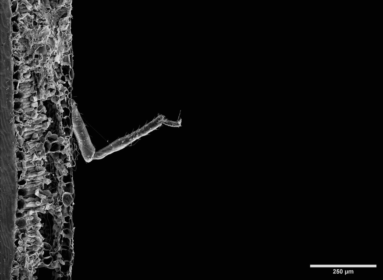
Plants are full of surprises, they are not only host for microorganisms but also for very small insects;

ImageJ=1.53f
unit=u00B5m

here we see a spider leg protruding from a cross-section of a banana leaf.

Specimen: Musa sikkimensis ( banana) leaf,  preparation and imaging from Anna Walkiewicz
Image: leaf cross-section with an unexpected inhabitant
Sample preparation: protocol for plant tissue preparation followed by Critical Point Drying (K850 CPD) and coating with 8nm of Pt (Q150V S plus).


Springtail-skin-1280x883.jpg

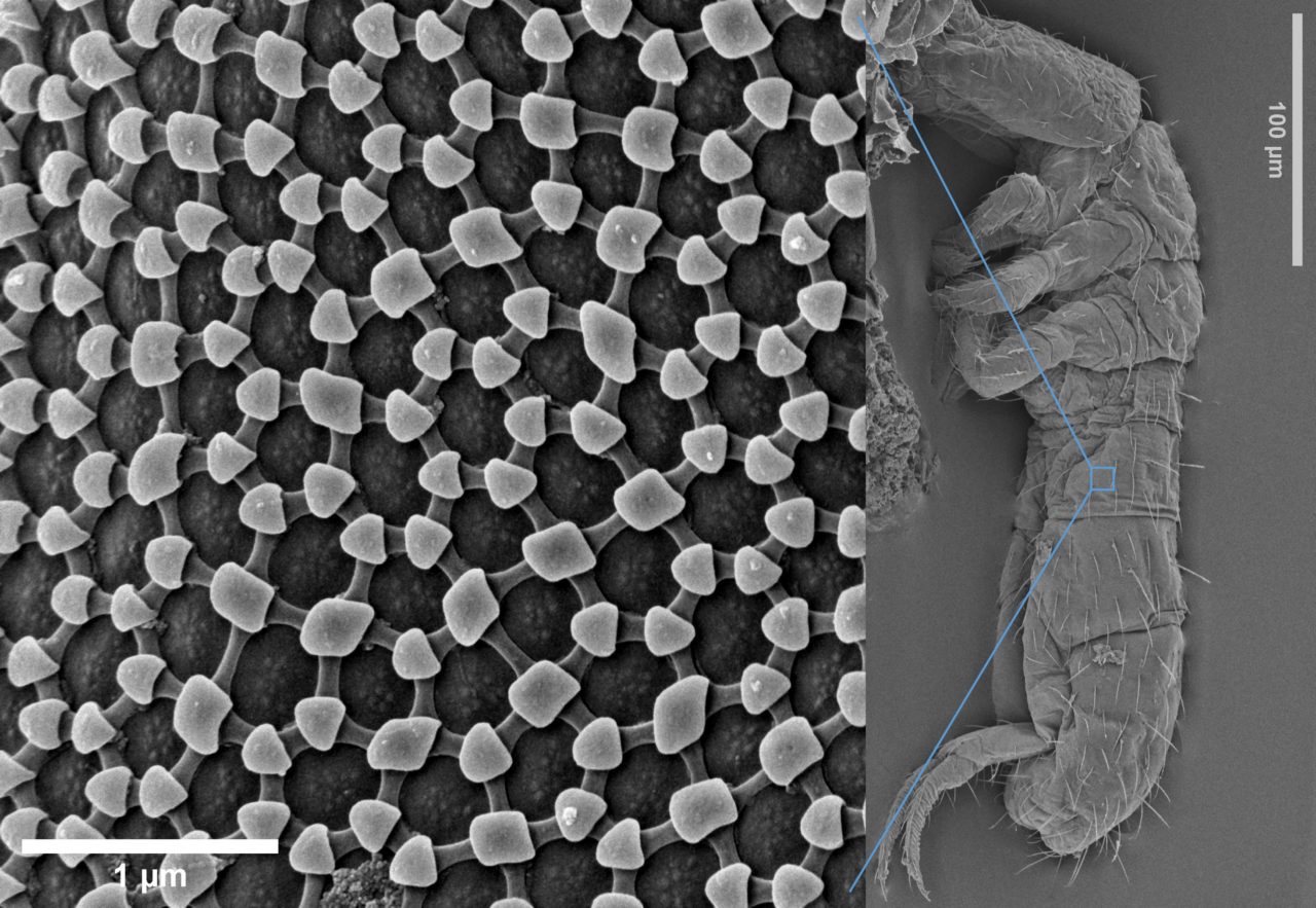
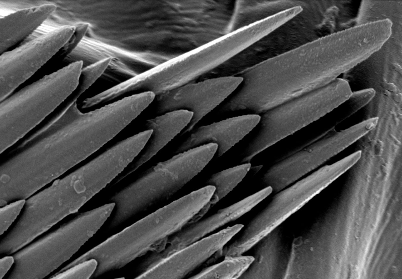
Springtails are extraordinary creatures, they are arthropods living in soil, plants and decaying material adapted to the environment by developing a special comb-like pattern on their skin. This rhombic-hexagonal structure is responsible for the non-wetting properties of the springtail body.

Sample: springtail colony from orchid bark. Sample preparation and imaging from Anna Walkieicz: bio preparation protocol followed by Critical Point Drying (K850 CPD) and coating with 8nm of Pt (Q150V S plus).


Picture1-1280x888.jpg

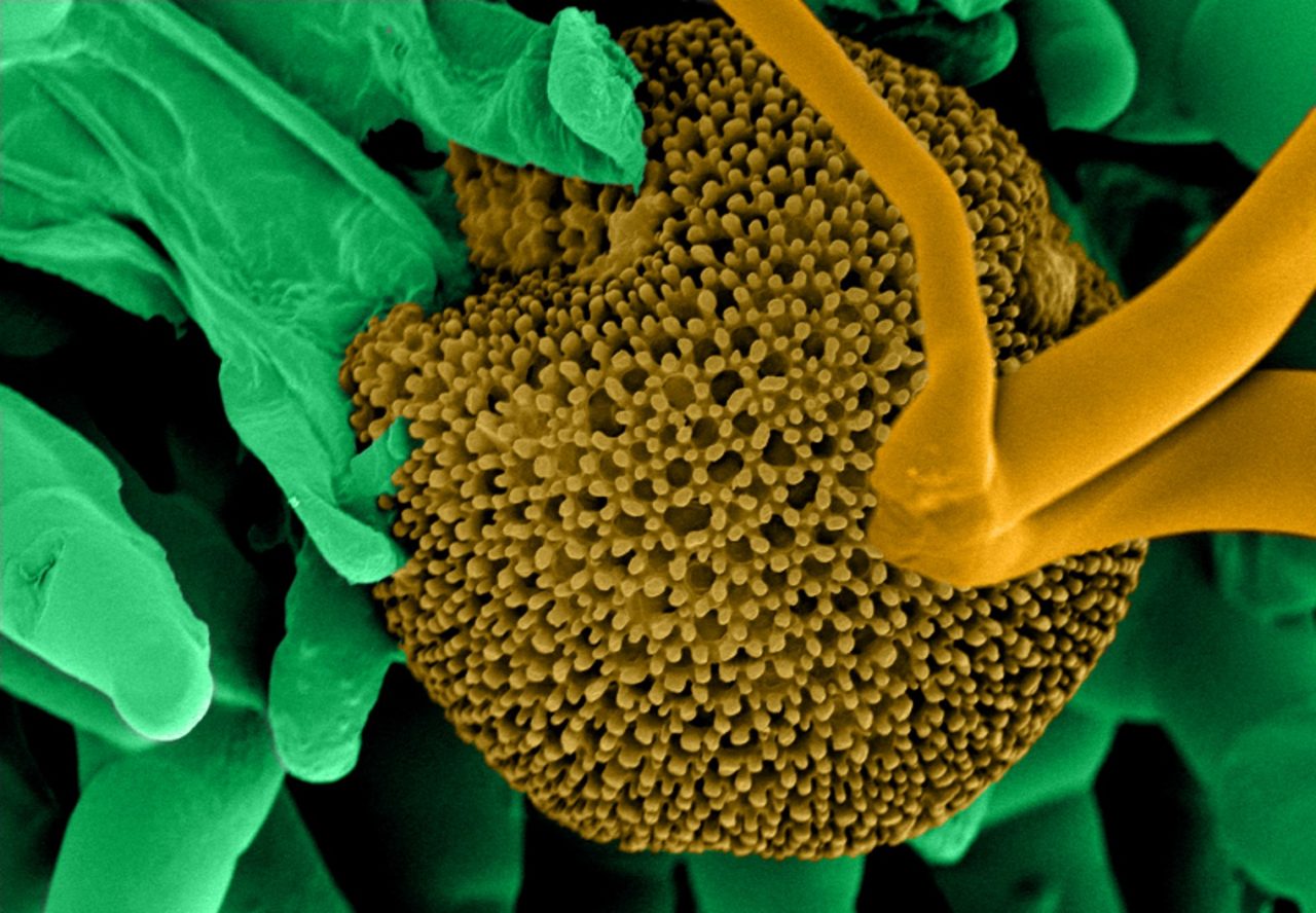
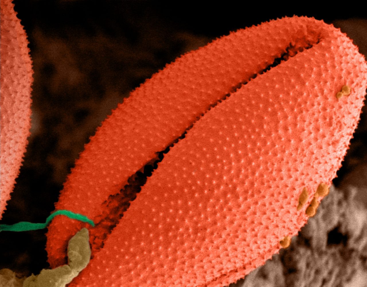
Pollen are small bioaerosol grains consisting of a single cell containing two male gametes essential for sexual reproduction of flowering plants. Typically, these bioaerosols range from 10 to 200 μm, are aerodynamic and low in density. These characteristics are essential for wind-pollination (anemophily) and/or insect-pollination (entomophily) – the transport of pollen from male stamen to the female stigma.

The grain wall is comprised of a tough chemically stable substance known as sporopollenin, which is highly resistant to environmental damage. It is punctuated with small openings called apertures which allows the male gametes to escape during pollination.

Pollen is typically classified by its distinctive structural features, i.e., variation in size, shape, number of apertures and surface texture, which also makes it possible to identify and classify a plant at family and genus level, and often even species level.

Anemophilous (inconspicuous or non-flowering) plants typically produce enormous quantities of very lightweight pollen grains, usually with air-sacs. Whereas entomophilous (insect-loving) plants produce pollen that is relatively heavy, prickly, and sticky to cling to the insects attracted by the flowers’ nectar and showy colours.

SEM micrographs of pollen grains below are from a Papaver Nudicaule (Iceland Poppy) and a Geranium (Wild Purple Cranesbill) plant image imaged using a FE-SEM prepared using the Quorum PP3010 cryo-preparation stage by Dr Mark S. Taylor.


Picture2-1280x889.jpg

Calcium oxalate is frequently found in plants, leafy vegetable such as spinach, almonds and dates in the form of tiny needle like raphides. Whilst these play a central role in controlling calcium regulation, herbivory and metal detoxification, unfortunately if consumed, calcium oxalate can lead to kidney stones. Out of all the 3 forms of calcium oxalate, the monohydrate form is the one widely reported to cause kidney problems.

Images show needle like raphides of calcium oxalate crystals observed at Quorum Technologies using an Ultra high-resolution Field Emission Scanning Electron Microscope (FE-SEM), equipped with a PP3010 automated, column-mounted, gas-cooled cryo-preparation/ transfer stage chamber www.quorumtech.com

Small cross-sections of the plants were cut and mounted on a universal cryo-stub using mounting media – a 50:50 mixture of Tissue-Tek OCT (Optimal Cutting Temperature) compound (Agar Scientific) and colloidal graphite (G303 Sakura) to fix and aid electrical conductivity.

The Tissue-Tek OCT (Optimal Cutting Temperature) being a mixture of clear water-soluble polyethylene glycol with polyvinyl alcohol and the colloidal graphite a mixture of graphite, propan‑2‑ol, butanol and 1-methoxy-2-propanol. The cryo-stub with sample was then mounted onto a specimen shuttle plunge frozen (vitrified) in slushed nitrogen (LN2) and transferred under vacuum conditions to the cryo-preparation chamber.

On the preparation cold stage, maintained at -140°C (anti-contaminator -175°C), the vitrified samples were cleaved with a cold knife, sublimated at -90°C for 2 – 3 min and then coated in situ with Iridium using a current of 5 mA for 60 – 90 s to ensure electrical conduction. Cryo-SEM imaging at -140°C (anti-contaminator -175°C) was carried out at acceleration voltages 1-1.5 kV and emission currents 15 – 20 µA at working distances 8 – 12 mm detecting secondary emitted electrons.
(Imaged by Dr Mark Taylor of Quorum Technologies, UK) #microscopy #Cryoprep #quorumtech #samplepreparation


cpd_water-daphnias_algies6nmPt_0023_SEUL-2-2XX.jpg

Quorum Technologies would like to celebrate International #womeninscienceday,
Dr Anna E. Walkiewicz is the Applications Specialist at Quorum Technologies. She holds a PhD from the University of Birmingham, where she researched the recognition of chirality at the nanoscale.

ImageJ=1.53f
unit=u00B5m

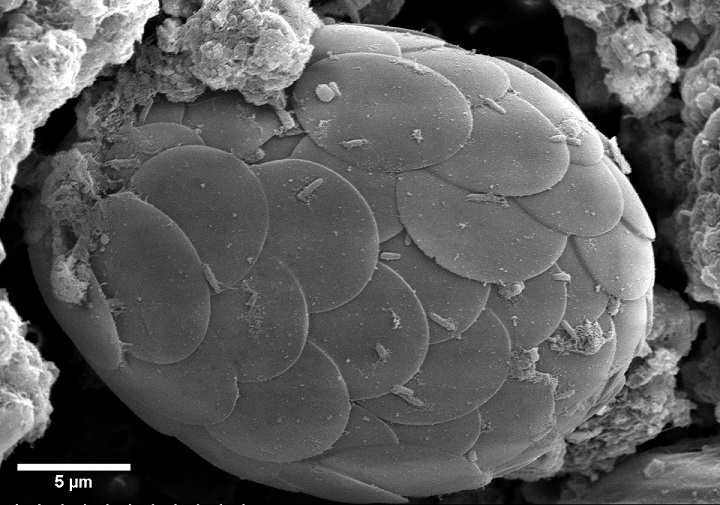
This is shelled ameboid from fresh water sample.
Sample preparation: Biological sample prep protocol followed by critical point drying in Quorum K850 and coated with 6nm of Pt in Quorum Tech Q150V S Plus. quorumtech.com


Diatoms-combined2Small-1280x896.jpg

Imaging Spotlight. 

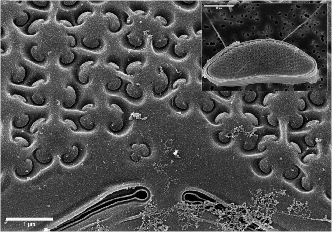
Diatom algae:
One of the most sophisticated frustule structures.
Sample preparation:
Biological tissue processing followed by critical point drying using Quorum Technologies K850 CPD and 8nm Pt coating in Q150VS Plus. Imaged by Dr Anna Walkiewicz, Applications Specialist
Daphnias algies


ImageForArticle_20774_16328329846236688.jpg

Electron microscopes use electrons rather than visible light to investigate and image samples, thus providing orders-of-magnitude enhancements on the magnification and resolution of traditional optical microscopes.

However, electron microscopes are much more complicated than optical microscopes, and the conditions that samples are subjected to are very difficult to work with.

SEM and TEM both depend on the transfer of electrons between a sample and the microscope itself, and thus imaging is performed under high vacuum. Moreover, samples must be conductive to enable electron transfer. This poses a difficulty for some sample types.

Although it is comparatively easy to use SEM or TEM to image solid metallic samples, for instance, imaging soft, hydrated and/or non-conductive samples — for example, biological tissues — can be challenging.

When left untreated, such samples lead to several issues. Moisture and gas from the sample easily evaporate inside the vacuum environment of an electron microscope sample chamber, thereby contaminating the microscope and damaging the sample.

Moreover, upon subjecting non-conductive samples to the incident electron beam within an electron microscope, the electrons do not conduct through the sample, making them accumulate in one place. This “charging” results in several imaging artifacts and can even make it impossible to image samples.

It is possible to solve these issues by the careful preparation of samples, specifically by sample coating. Using a layer of conductive material (carbon or metal) to coat samples has several purposes. There are two key reasons for coating: to make the sample conductive, which avoids “charging” effects, and to encapsulate samples to eliminate off-gassing or evaporation.1

Specifically, metallic coatings can facilitate better thermal conductivity, safeguarding the sample from heat damage from the incident electron beam. They can even localize the signal to the actual surface of the sample, thus enhancing the signal-to-noise ratio and secondary electron emission.

Carbon coatings offer certain distinct advantages. Carbon coatings for electron microscopy are amorphous, conductive layers transparent to electrons. This implies that carbon coatings are particularly valuable for making non-conductive samples amenable to energy-dispersive x-ray spectroscopy (EDS).2

Achieving High-Quality Carbon Coatings

For SEM and TEM applications, metallic coatings such as tungsten and gold can be done through sputtering. However, the same is not true of carbon. Although carbon can be sputter-coated, the resulting coatings exhibit high hydrogen concentrations, which makes carbon sputtering unsuitable for electron microscopy applications.

Alternatively, high-quality carbon coatings can be performed through thermal evaporation of carbon in vacuum. Two similar techniques can be used to achieve this — using carbon fiber or using a carbon rod.

In the carbon rod coating method, two carbon rods with a sharpened contact point between them are used. This is also called the Brandley method.

The process involves passing current between the two rods, ensuring very high current density at the sharpened contact point, which leads to very high levels of resistive heating. This causes the evaporation of carbon from the surface. This can be achieved with a ramped current or a pulsed current.

In the carbon fiber technique, a carbon fiber is mounted between two clamps, and a pulsed current is passed along it. This leads the carbon to evaporate from the surface of the fiber.

Both techniques exhibit unique differences in quality. The carbon fiber technique facilitates certain control over coating thickness by tweaking the number of current pulses and a pulse length. This makes it appropriate for TEM grid applications and analytical SEM applications like EDS and electron backscatter diffraction (EBSD). Yet, pulsed carbon fiber coatings essentially contain higher levels of debris.

Carbon rod coatings are “cleaner” and of better quality. Carbon rod coatings made in high vacuum with ramped current offer the maximum quality coatings, suitable for high-resolution TEM applications and crucial SEM applications.

In its pulsed version, the technique can be employed to achieve thicker coatings for SEM, particularly for wavelength-dispersive X-ray spectroscopy (WDS) and EBSD. In such applications, it is important to select carbon rods with the maximum purity to achieve the maximum possible coating quality.

Carbon Coating Solutions from Quorum Technologies

Quorum Technologies has come up with the new Q Plus Series that offers an all-in-one solution to attain high-quality carbon coatings for all electron microscopy applications.

With the ability to coat both carbon fiber/cord and carbon rod, the carbon evaporators employ easy-change inserts to allow users to easily switch between the two modes.

The new Q150V Plus from Quorum Technologies offers the highest vacuum of 1 x 10⁻⁶ bar for superior results. The lower background pressure implies oxygen, nitrogen and water vapor are eliminated from the coating chamber, restricting chemical reactions while executing the coating process. This leads to impurities or defects. Lower scattering also implies amorphous carbon films of higher purity and high density.

References

  1. Goldstein, J. I. et al. Coating Techniques for SEM and Microanalysis. in Scanning Electron Microscopy and X-Ray Microanalysis: A Text for Biologist, Materials Scientist, and Geologists (eds. Goldstein, J. I. et al.) 461–494 (Springer US, 1981). doi:10.1007/978-1-4613-3273-2_10.
  2. Heu, R., Shahbazmohamadi, S., Yorston, J. & Capeder, P. Target Material Selection for Sputter Coating of SEM Samples. Microscopy Today 27, 32–36 (2019).
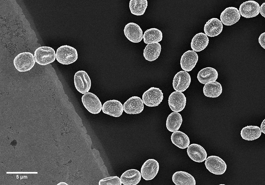
  3. Image shown – Fungi spores on a TEM grid (Cu, 300 mesh with 5nm carbon film produced with Q150VES Plus coater). Image Credit: Quorum Technologies

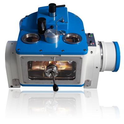
pp3000T.jpg

Quorum’s PP3000T has now reached end-of-life support.

Please be aware if you have one still installed you will no longer be able to receive parts or service support from Quorum Technologies Ltd.

If you have any queries, please don’t hesitate to contact the sales team at [email protected]

All products currently available, in support, or no longer supported can be found on our website

https://www.quorumtech.com/products-and-parts-supported/


Screenshot-2020-08-27-at-09.29.07-e1599595360963.png


To all our customers, suppliers, and partners,

I hope that you, your family and friends are safe and well in these unprecedented times. I wanted to update you concerning the measures that Quorum Technologies has put in place to counter the impact of the pandemic on our business and how we are continuing to support users of Quorum equipment.

Quorum Technologies remained open for business throughout the pandemic, thanks to the flexibility and cooperation of our staff and the continued support of our customers and suppliers. Thank you for helping us to make it this far.

One of the reasons we felt it essential to keep operating was that our sample preparation equipment for Electron Microscopes was seen as a key technology for essential medical research. I am proud to say that, during the lockdown, we provided support and equipment to customers who are directly involved in research into the SARS-CoV-2 coronavirus and hopefully contributed in a small way towards eventually beating this virus.

Communication during the pandemic:

Our production sites in Laughton and Lewes were closed to external visitors at the start of lockdown. We have now opened again, with some restrictions:

  • All visits are by appointment only.
  • Instead of visiting the factory, your contact at Quorum may suggest off-site alternatives.
  • If you do come to the factory, you will be required to comply with social-distancing measures, wear a face-mask, and follow strict guidelines on handwashing and sanitisation of work areas.

Many of our staff were directed to work from home, in keeping with the instructions issued by the U.K. government, or were furloughed during the lockdown. We have reorganised our offices and put in place additional measures to comply with guidelines for a CoViD-secure workplace and are now recalling staff to full-time work. Some back-office staff will continue to work from home but will be contactable as normal.

When contacting Quorum by phone, please use the main switchboard number, +44 (0) 1323 810981, and your call will be redirected to the appropriate number.

While we have been lucky in that no one at Quorum has yet contracted CoViD-19, the virus is still circulating in the U.K., and we may have unplanned absences due to illness or quarantine. For that reason, we cannot guarantee that individual staff members will always be available, and we continue to ask that you direct your calls and emails to teams, rather than to individuals, to enable a prompt response.

Our team emails are:

Accounts receivable/payable:         [email protected] 

Procurement:                                   [email protected]

Product sales:                                  [email protected]

Technical support:                            [email protected]

Supply of goods

Our Purchasing team has been working closely with our suppliers to ensure a continued supply of key parts, but there has been some disruption in supply chains which has impacted some of our older instruments. We are currently unable to guarantee lead-times for the following products:

K975X/K975S Turbo-Pumped Thermal Evaporators

K750X Peltier-Cooled EM Freeze Dryer

K775X Liquid Nitrogen Cooled Turbo-Pumped EM Freeze Dryer

K1050X RF Plasma Etcher/Asher/Cleaner

Customers with an urgent need to guarantee supply should contact the sales team as soon as possible to discuss. You can reach our sales team via the main switchboard number or by emailing them at [email protected].

Field service and installations

We have resumed field service activities, subject to complying with CoViD-secure guidelines. If you want to schedule a visit by our service engineers, you will be asked to provide information on the measures in place at your facility so we can conduct a risk assessment before scheduling a visit.

Unfortunately, our ability to travel overseas continues to be constrained by travel bans in certain countries and by the need to comply with quarantine requirements in others. We recognise that our equipment can be vital to research and we will travel where we can, but it may not be possible in every case. For example, if your country has a prohibition on visitors from the U.K. or requires an extended period of quarantine on arrival, we would be unable to schedule a field visit. Where travel is not possible, we will do our best to offer remote support by phone or email.

You can reach our service team via the main switchboard number or by emailing them at [email protected].

Thank you again for your support throughout this difficult period. We are, more than ever, grateful for that support and proud of the fact that you choose to work with us. I will update you again if we make further changes but please do not hesitate to contact me in the meantime if you have any concerns or questions.

Thank you for working with Quorum Technologies.

Yours sincerely,

Tony Larkin

Managing Director
Quorum Technologies Ltd


UNITED KINGDOM

Headquarters

Judges House, Lewes Road,
Laughton, East Sussex.
BN8 6BN

+44 1323 810981

www.quorumtech.com

[email protected]

UNITED KINGDOM

Working Hours

8.30 – 5.00 pm Mon – Thu
8.30 – 4.00 pm Fri
QUICK LOOK

Latest News


No more posts


GET IN TOUCH

Follow Our Socials

Keeping in contact with our customers via social platforms as well as traditional telephone and email.