Electron Microscopy Archives - Quorum Technologies Ltd

Front-Cover-Coffee-Talks_-002-1280x688.jpg

In this series Dr Anna Walkiewicz, the Quorum Application Specialist, will talk about different processes within the sample preparation workflow and breaking them down into simple steps, solving common problems and looking at how to improve efficiency and efficacy.

Coating for SEM. How to choose vacuum level, metal and thickness.

In this Coffee Talk Dr Anna Walkiewicz will guide you through metal coating for SEM imaging. During the tutorial, you will learn what metal to choose for different applications and how coating process parameters influence coating quality.

Register here


ct-anna-1280x363.jpg

Coating for SEM. How to choose vacuum level, metal and thickness.

In this Coffee Talk Dr Anna Walkiewicz will guide you through metal coating for SEM imaging. During the tutorial, you will learn what metal to choose for different applications and how coating process parameters influence coating quality.

You can watch the complete series on our website soon.


Register for Mounting samples and coating; how to do it correctly 

    I consent to Quorum Technologies collecting and storing my personal information for further marketing.I DO NOT consent to Quorum Technologies collecting and storing my personal information for further marketing.


    CT04-sample-mounting-1280x1540.jpg

    In this Coffee Talk you will learn about techniques used to attach samples to SEM stubs, different materials and accessories that could be used to expose parts of interest in the best way. Dr Walkiewicz will offer tips for different sample types and explain the method in which you mount could affect the efficacy of coating.

    Upcoming episodes

    • 9th November @11am GMT CT 05 – Coating for SEM. What do we use for coating and when? Vacuum level, metal choice, thickness.

    You can also watch all previous episodes on our website.


    Register for Mounting samples and coating; how to do it correctly 

      I consent to Quorum Technologies collecting and storing my personal information for further marketing.I DO NOT consent to Quorum Technologies collecting and storing my personal information for further marketing.


      CPD_-Image-1280x1381.jpg

      09/27/2022by Sarah Hall

      In this series of mini-talks, Dr Anna Walkiewicz will be describing the process of sample preparation by breaking it down into simple steps, aiming to solve common problems, while looking at how to improve efficiency and efficacy.

      In the third episode of the Coffee Talks series, Dr Anna Walkiewicz will guide you through the process of Critical Point Drying.

      You will learn what kind of samples could be dried this way and how to prepare them beforehand to achieve the best outcome. Dr Anna Walkiewicz will also highlight potential issues and give advice on how best to address them.

      Upcoming episodes

      • 26th October @11am BST CT 04 – Mounting samples – exposing the most relevant parts. Coating – why do we coat samples?
      • 9th November @11am GMT CT 05 – Coating for SEM. What do we use for coating and when? Vacuum level, metal choice, thickness.


      Register for Critical Point Drying: A guided tour through a complex process below: 

        I consent to Quorum Technologies collecting and storing my personal information for further marketing.I DO NOT consent to Quorum Technologies collecting and storing my personal information for further marketing.


        marshmallow-1280x800.jpg

        In this series Dr Anna Walkiewicz, the Quorum Application Specialist, will talk about different processes within the sample preparation workflow and breaking them down into simple steps, solving common problems and looking at how to improve efficiency and efficacy.

        The first coffee talk, The Scary Vacuum, will be addressing issues regarding the vacuum: why do we need to consider if our samples are sensitive to the change of pressure and how they will behave in the vacuum environment.  Dr Anna Walkiewicz will be giving advice how to prepare samples in the correct way, so we see their natural appearance rather than an outcome of the pressure change modifying their morphology. Register here Coffee Talks with Dr Anna Walkiewicz – The Scary Vacuum 14th September – Quorum Technologies Ltd


        PitcherplantLeaftop_002scale-1280x848.jpg

        See a piece below from our customers at the Cambridge Advanced Imaging Centre at Cambridge University.

        Cambridge Advanced Imaging Centre (CAIC) forms the hub of a network that draws together imaging activity across the whole University to serve the biomedical community. Many powerful modern imaging techniques have recently been developed, yet their exploitation in biology and biomedical research has largely been prevented by the high cost of their translation into widely available instruments and the specialist skills required for their use, including handling the vast volumes of data they generate. As a consequence, instruments developed in laboratories within specific Cambridge research programmes are often not available to the University’s wider biomedical research community.

        Our Verios 460 SEM is equipped with a Quorum PP3010T cryo-SEM preparation system. Cryo-SEM allows imaging of soft, hydrated biological or materials samples in a frozen state. One of the major advantages of cryo-SEM is that samples can be imaged with minimal preparation without the need of any prior fixation or dehydration steps; a disadvantage is that sample throughput is fairly low compared to other methods. Specimens are mounted on an appropriate cryo-SEM shuttle, are plunge-frozen in slushed liquid nitrogen and then transferred to a cooled prep stage. Once on the prep stage, samples can be fractured, sublimed to remove any residual ice, and coated to improve contrast and conductivity (CAIC routinely uses a platinum target). Then, the sample is transferred onto the SEM cryo-stage for imaging. As cryo-SEM requires about 2h of run-up and run-down time of the system apart from the actual imaging time, we recommend half-day or whole-day bookings.

        Some images below from their cryo-SEM


        Springtail-skin-1-1280x883.jpg

        In this series of mini-talks, Dr Walkiewicz will be describing sample preparation process and breaking it down into simple steps, solving common problems and looking at how to improve efficiency and efficacy.

        In this second episode of the series, Dr Walkiewicz will be looking at dehydration of samples, why it is so important, why we dehydrate, do we need anything before dehydration, and means of tissue fixation.

        Upcoming episodes

        • 10th October @11am BST CT 03 – Critical Point Drying – is it really so complex?
        • 24th October @11am BST CT 04 – Mounting samples – exposing the most relevant parts. Coating – why do we coat samples?
        • 7th November @11am BST CT 05 – Coating for SEM. What do we use for coating and when? Vacuum level, metal choice, thickness.

        Register for the above talks on the specific event page


          D12650-008-scaled-e1657786179383-1280x1120.jpg

          Dr Anna Walkiewicz, the Quorum Technologies Application Specialist, is running a series of informative and educational webinars across this autumn looking at all aspects of sample preparation. The first talk is on 12th September at 11am BST, until 11.30am BST.

          In this series, Dr Walkiewicz will be tackling different processes within the sample preparation process and breaking it down into simple steps, solving common problems and looking at how to improve efficiency and efficacy.

          • CT 01-The Scary Vacuum – 12th September – How will samples behave in the vacuum?
          • CT 02-Dehydration 26th September – Why is it so important? Why do we dehydrate samples? Do we need anything before dehydration? Means of tissue fixation
          • CT 03- Critical Point Drying 10th October – is it really so complex?
          • CT 04- Mounting samples 24th October – exposing the most relevant parts. Coating – why do we coat samples?
          • CT 05- Coating for SEM 7th Nov – What do we use for coating and when? Vacuum level, metal choice, thickness

          To register to watch these events, please visit our events page and fill out the form


          Final.jpg

          Cryo-SEM of microcapsules
          Omega-3 polyunsaturated fatty acids (ω-3 PUFAs) are extremely important in human physiology. The primary source is typically obtained from fish oils. Whilst there is scientific evidence linking the consumption of ω-3 PUFAs to reduced risk of breast and prostate cancer, high blood pressure and blood circulation – the daily intake in many Western societies falls below the recommended minimum levels. Taking supplements and enrichment of foods is an ideal way to increase the ω-3 PUFAs levels in the diet.

          Microencapsulation with modified cellulose by spray-drying provides an alternative way of supplementing where the final product is a fish oil powder additive. The cellulose coating surrounding the oil also improved the stability against oxidative rancidity for up to 12 – 24 months.

          The microstructure and encapsulation efficiency can be determined by various methods including cryo-electron microscopy. The cryo-SEM micrograph below shows a variety of spray-dried microcapsules ranging from 1.5 to 17 μm, prepared for imaging using the Quorum PP3010. Preparation includes applying a thin layer on mounting media, vitrification in slushed N2 (-210 °C) and sputtering with Pt (1 – 2 nm). Inset image shows the cellulose coating of a fractured capsule of diameter ≈ 25 μm with wall coating thickness ≈ 571 nm.

          Dr M. S. Taylor


          crystals_2nmAuPd_V_0058_mag100k.jpg

          Electron microscopy techniques rely on the transfer of electrons between sample and microscope. For conductive samples, this is easily achieved – however, non-conductive, or poorly conducting samples must be coated with an electrically conductive coating to produce usable images. A high-quality coating is essential to obtain high-quality images. Quorum Technologies developed the Q Plus Series to provide researchers with a versatile and high-performance coating to rival major manufacturers, without the associated price tag.  

          The Role of Coatings in Electron Microscopy

          Scanning Electron Microscopes (SEM) and Transmission Electron Microscopes (TEM) work similarly to Optical Microscopes but rather than probing materials using light, they use electrons. Optical microscopes are diffraction-limited (to a maximum resolution of about 200 nm) whereas electron microscopes can produce beams of electrons with much smaller wavelenght1 and surpass the resolving power of optical microscopes by several orders of magnitude. As a result, they are the most powerful microscopy techniques in the world.

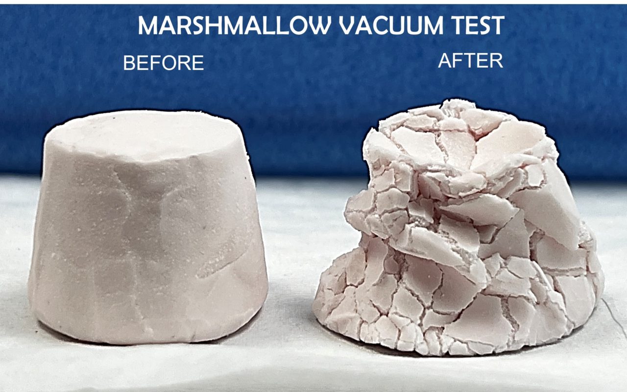
          Using electrons instead of light, however, introduces other complications. Both techniques (SEM and TEM) rely on the transfer of electrons between sample and microscope; therefore, it can be difficult (or near impossible) to obtain a usable image signal from samples with poor or no conductivity. This is especially true of SEM, where samples are bombarded with an electron beam: poorly conductive or non-conductive samples will rapidly accumulate charge under these conditions, leading to image distortion as well as thermal and radiation damage to the sample. In extreme cases, the sample may accumulate sufficient charge to decelerate the primary beam, acting as an “electron mirror” and preventing an image altogether.2

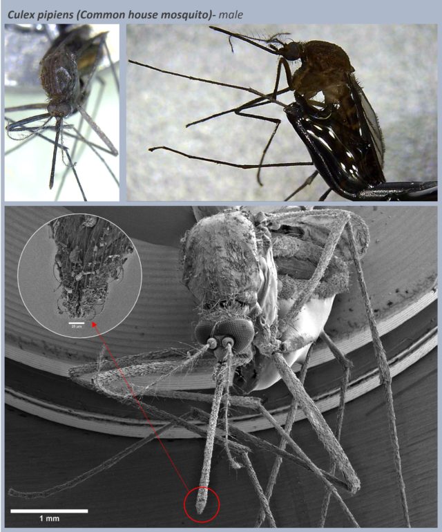
          To overcome this, poorly conducting samples are coated with a thin layer of metal or carbon. This makes the surface conductive, eliminating charge accumulation and enabling a better signal to be obtained by the microscope. Coating techniques are widely used for imaging biological or organic samples since these are typically non-conductive and easily damaged by the electron beam.

          While the primary role of coating in SEM is to increase electrical conductivity and prevent “charging”, it also has several other useful effects:

          • Coating a sample with a thermally conductive material such as gold, silver, copper, aluminium can reduce thermal damage from the primary electron beam.
          • Particulate matter and fragile organic samples can be mechanically stabilized and held in place by a thin layer of carbon.
          • Coating organic samples that contain trapped gas or moisture protects both sample and microscope from being contaminated by off-gassing.
          • Metallic coatings can be used to minimize the volume of penetration of the electron beam, localizing scanning to the very surface of a sample. This can also dramatically increase the emission of secondary and backscattered electrons.

          Download our Guide to Coating for Electron Microscopy Here


          The Impact of Coating Quality

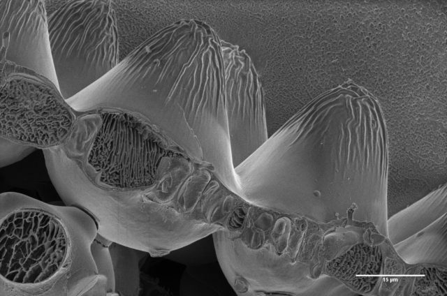
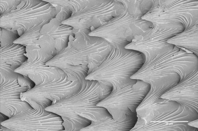
          When working with a coated sample in an electron microscope, it is the coating itself that gets directly imaged. The quality of the coating, therefore, places a hard limit on the quality of the images that can be obtained.

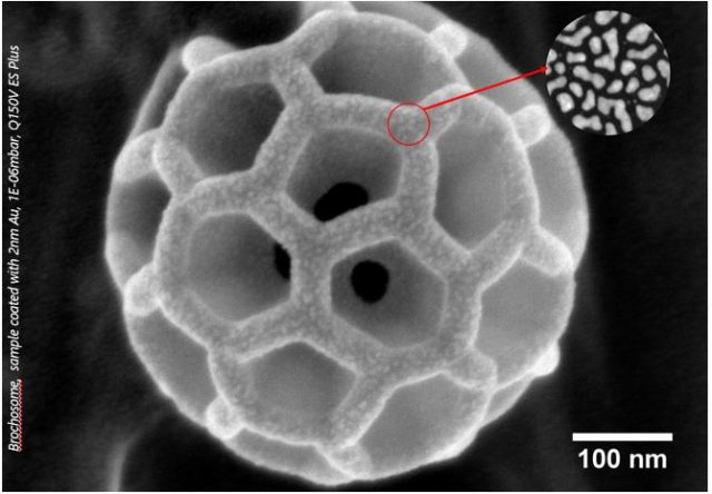
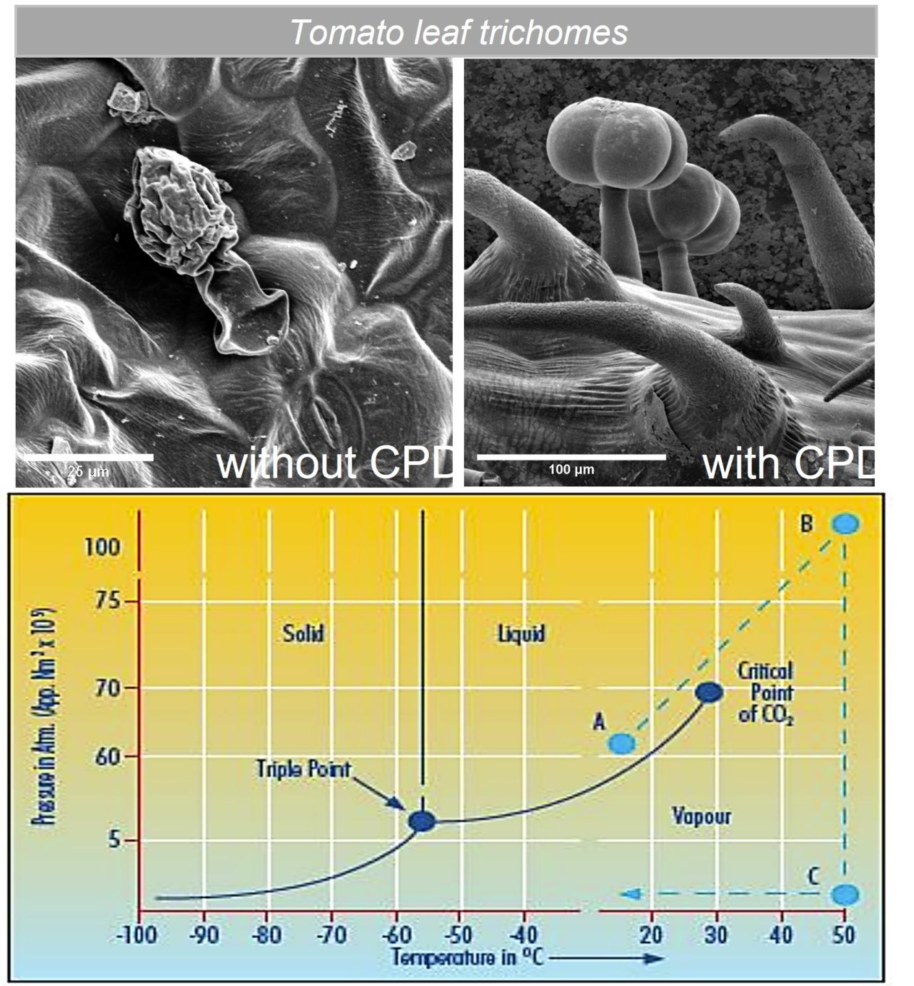
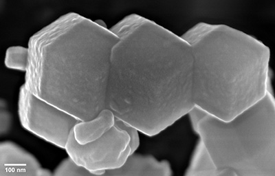
          When imaging very small structures (such as electrospinning fibres doped with copper nanocrystals), depositing a coating that is too thick can easily bury meaningful information. It is vital that coating thickness can be precisely controlled and tailored to the features that are being interrogated.3

          In the worst cases, poor quality coating equipment introduces contamination and can irreparably damage samples. Researchers often opt for cheap coaters to save money, only to find that their costs increase due to additional microscope time and ruined samples.

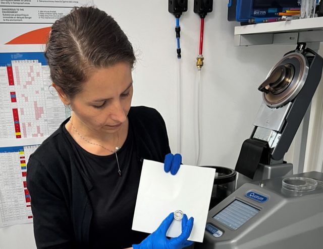
          However, thanks to the Q Plus Series from Quorum Technologies, it is no longer necessary to pay a premium to obtain state-of-the-art coatings.

          The Q Plus Series: Affordable and High-Quality Coating

          The Q Plus Series is the latest iteration of Quorum’s world-leading range of coaters; offering cutting-edge sputter and evaporation coating in a single easy-to-use platform. Quorum’s turbomolecular-pumped coaters are suitable for both oxidizing and non-oxidizing metals, while our low-cost rotary-pumped sputter coaters are suitable for non-oxidizing metals. The Q Plus Series is suitable for sputter coating and evaporating carbon coating for SEM, FE-SEM and TEM applications.

          This new range of coaters is designed to enable researchers to exercise precise control over coating thickness, whatever their application requirements. For the highest level of performance, the Q150V Plus provides an ultimate vacuum of 10-6 mbar; removing oxygen, nitrogen and water vapour from the chamber and eliminating chemical reactions during the sputtering process. The Q150V Plus also enables the production of finer grain size and thinner coating for ultra-high-resolution applications (beyond 200,000x magnification). Low scattering enables the formation of high-purity amorphous carbon films of high density.

          All models in the Q Plus Series feature a touch-screen interface as well as status LEDs and audio notifications for straightforward and intuitive control. Integrated 16 GB memory allows the storage of over 1000 recipes to be stored, and a USB port enables upgrades and downloads of log files.

          To find out more about the Q Plus Series of coaters, view our brochure or get in touch with us today.

          To view our latest webinars on coating technologies, we invite you to view our series here:
          1. Practical advice in sample preparation for SEM
          2. How to achieve appropriate metal coating quality for my application
          3. Carbon coatings and Glow Discharge for TEM


          References and Further Reading

          1. The Diffraction Barrier in Optical Microscopy. Nikon’s MicroscopyU https://www.microscopyu.com/techniques/super-resolution/the-diffraction-barrier-in-optical-microscopy.
          2. Goldstein, J. I. et al. Coating Techniques for SEM and Microanalysis. in Scanning Electron Microscopy and X-Ray Microanalysis: A Text for Biologist, Materials Scientist, and Geologists (eds. Goldstein, J. I. et al.) 461–494 (Springer US, 1981). doi:10.1007/978-1-4613-3273-2_10.
          3. Ahire, J. J., Neveling, D. P. & Dicks, L. M. T. Polyacrylonitrile (PAN) nanofibres spun with copper nanoparticles: an anti-Escherichia coli membrane for water treatment. Appl Microbiol Biotechnol 102, 7171–7181 (2018).


          UNITED KINGDOM

          Headquarters

          Judges House, Lewes Road,
          Laughton, East Sussex.
          BN8 6BN

          +44 1323 810981

          www.quorumtech.com

          [email protected]

          UNITED KINGDOM

          Working Hours

          8.30 – 5.00 pm Mon – Thu
          8.30 – 4.00 pm Fri
          QUICK LOOK

          Latest News


          No more posts


          GET IN TOUCH

          Follow Our Socials

          Keeping in contact with our customers via social platforms as well as traditional telephone and email.